I interpret tissue findings alongside dose, exposure and the wider study data to assess treatment relationship, adversity and reversibility. My experience spans small molecules, oligonucleotides and biologics.
Study pathologist
Primary evaluation
Examine the agreed tissues and record diagnoses, distribution and severity.
Integrate gross findings, organ weights, clinical pathology and relevant study data.
Prepare the pathology report and discuss any follow-up assessment.
Independent review
Pathology peer review
An independent review of the study pathologist’s diagnoses and interpretation, with the scope agreed in advance.
Define organs, groups, slides and records; identify report and data versions.
Review diagnoses, grading, terminology and interpretation.
Discuss differences with the study pathologist. Provide a signed and dated peer-review statement identifying the scope, source versions, conclusions and any remaining differences.
Particular experience
Oligonucleotide safety and neuropathology
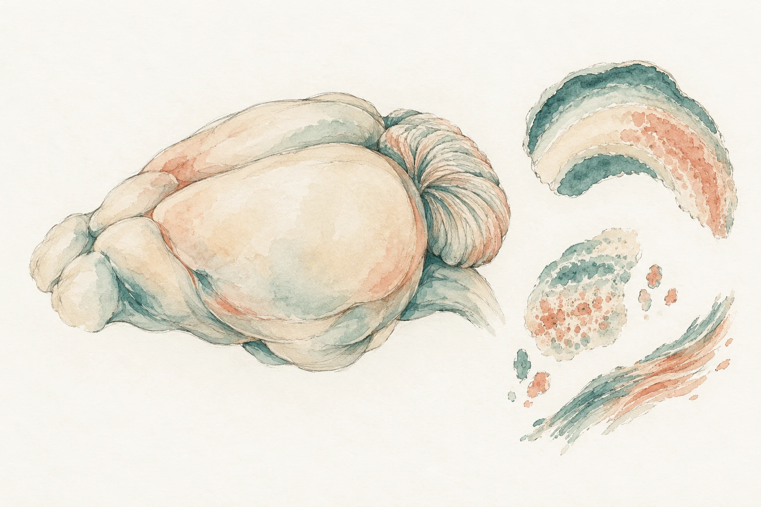
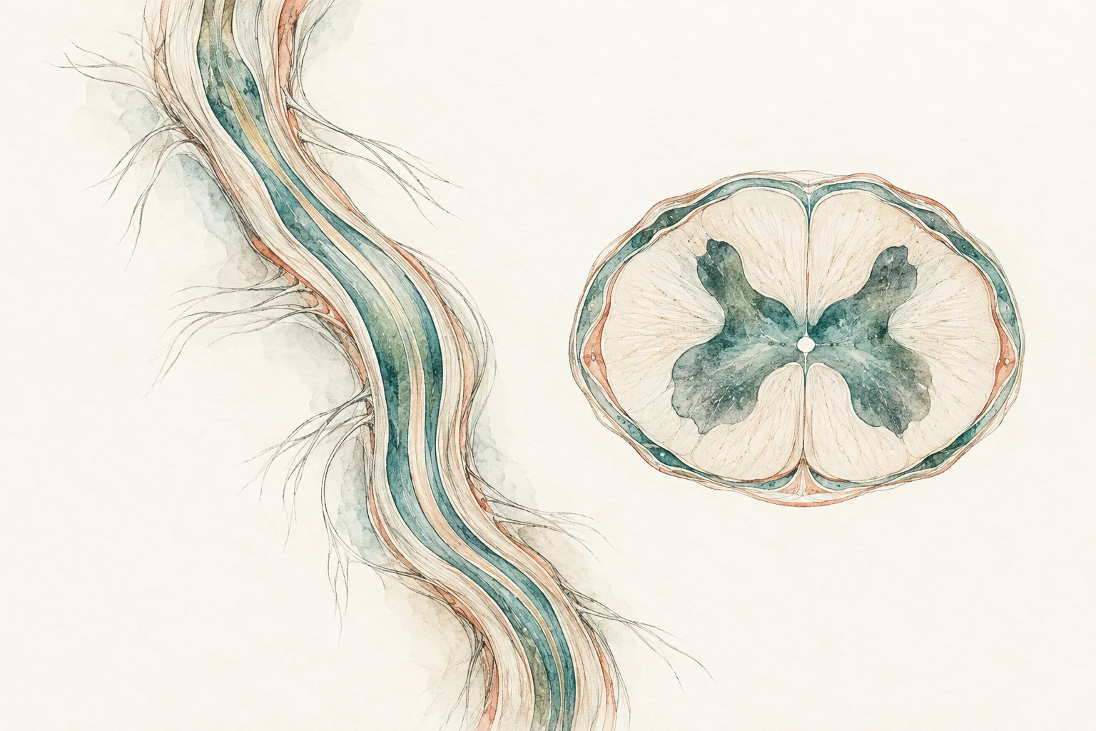
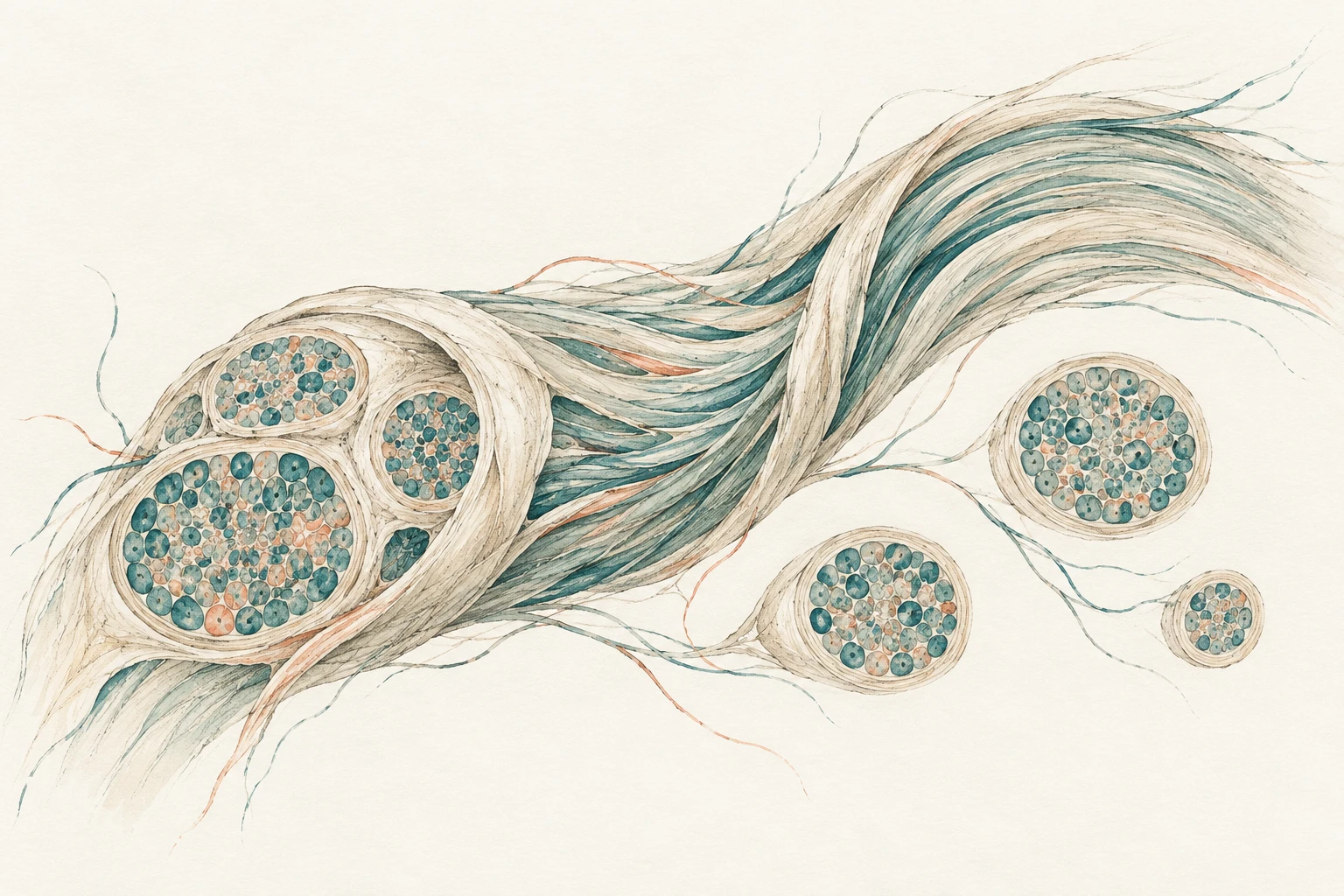
My neuropathology work spans ASO and siRNA safety studies, including intrathecal programmes. I assess the brain, spinal cord, dorsal root ganglia and peripheral nerves.
Assessment and ancillary preparations
I consider lesion distribution alongside administration route, clinical observations and tissue quality. H&E can be supported by IBA-1, calbindin, GFAP and Fluoro-Jade preparations where available.
BrainSpinal cordPeripheral nerve
Planning the pathology
Pathology input into study design
I help the study team plan the pathology assessment:
Tissue selection, sampling, trimming and orientation.
Necropsy and recovery time points.
Terminology, grading, blinding and ancillary endpoints.
Primary read, peer review and reporting schedule.
Dose-range-finding and MTD studies
Characterise target-organ findings and dose-related tissue responses to inform dose selection and follow-up assessment.
IND-enabling safety studies
Single- and repeat-dose programmes, with tissue findings interpreted alongside dose, exposure, duration and other study observations.
Recovery assessments
Comparison of findings at treatment-end and recovery time points to assess their persistence, progression or reversibility.
Organ-focused investigations
More detailed assessment of a particular organ or lesion, using additional levels, stains or defined measurements where appropriate.
Study-specific arrangements
GLP and non-GLP studies
My experience includes primary evaluations and peer reviews of GLP and non-GLP studies. For a GLP assignment, the arrangements are defined with the responsible test facility and Study Director.
The study pathologist is responsible for the final pathology evaluation and report. The Study Director retains overall responsibility for the GLP study.
Role and procedures
Responsibilities, examination scope, applicable SOPs and the versions of the protocol and records to use.
Data and quality assurance
Agreed data-capture systems, review documentation and quality-assurance arrangements.
Reporting and retained records
Report or peer-review statement, handling of changes and transfer or retention of study records.
Ancillary work
The GLP or non-GLP status of additional stains, exploratory measurements and image analyses is specified for each component.
The approach to histopathology peer review is informed by OECD guidance and FDA guidance, as applicable to the study.
Material and deliverables
Agree the working arrangements.
I work with multiple CROs and use Patholytix, Provantis and Pristima, adapting to the study team’s data-capture and reporting workflow. Before examination, we agree the slide or image inventory, supporting records and system access.
You receive a pathology report, peer-review statement or focused interpretation, according to the commissioned role.
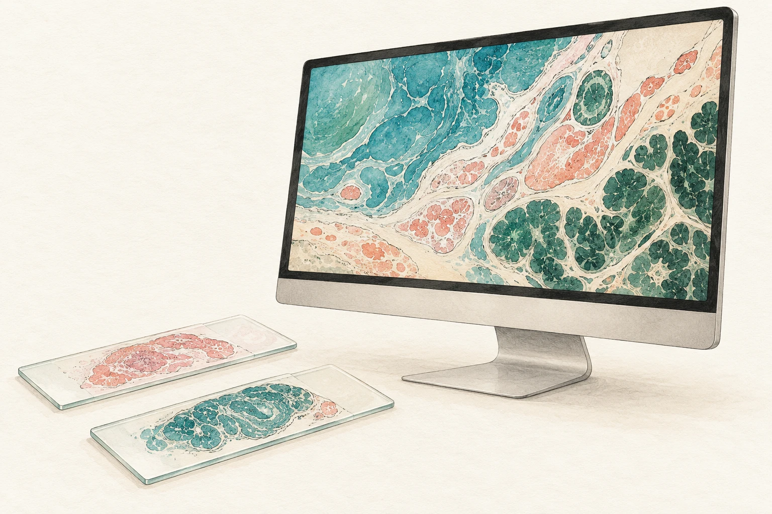
Digital review and whole-slide images
Before examination, we confirm image suitability, tissue coverage, viewing arrangements and documentation. Study-specific procedures address the use and retention of the images. FDA whole-slide imaging guidance (2023) informs these arrangements where applicable.