Investigative pathology

Pathology for
animal models

Histology, lesion scoring and tissue measurements, planned around the biological question.

Applications include neuroinflammation and demyelination, neurodegeneration and proteinopathies, cancer and immuno-oncology, MASH and hepatic fibrosis, diabetes and metabolic disease, IBD and colitis, inflammatory arthritis, pulmonary fibrosis, kidney injury and chronic kidney disease, osteoarthritis, inflammatory skin disease, allergic airway disease and asthma, myocardial infarction and cardiac fibrosis, atherosclerosis, wound healing and tissue regeneration, retinal disease and ocular neovascularisation, muscular dystrophy, ovarian and PCOS models, among others.

The examples below explore the pathology approach in more detail. Selected projects from my completed work are described on the Experience page.

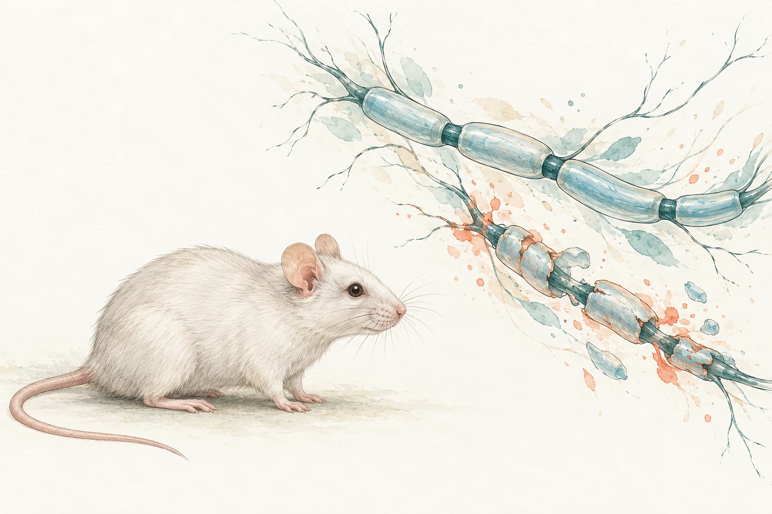
Editorial illustration of a laboratory mouse beside separate flowing axon and myelin motifs.
Mouse model · neuroinflammation and myelin

Model application

Neuroinflammation and demyelination

MOG-induced EAE in mice

Histology separates inflammatory infiltrates, myelin damage and axonal injury. Read alongside the clinical disease course, these endpoints help characterise the tissue response to treatment.

Endpoints, sampling and interpretation
Pathology endpoints
Inflammatory distribution and severity, the extent of demyelination, and axonal injury where appropriate. H&E, myelin stains and selected axonal or glial markers answer different parts of the question.
Sampling
Agree spinal-cord levels, relevant brain regions and disease stage before scoring. Lesion distribution changes over time; anatomical levels and tissue quality need to be comparable between animals.
Interpretation
Reduced inflammation does not by itself establish myelin repair. A remyelination claim needs methods that can substantiate it.
Related expertise: safety neuropathology
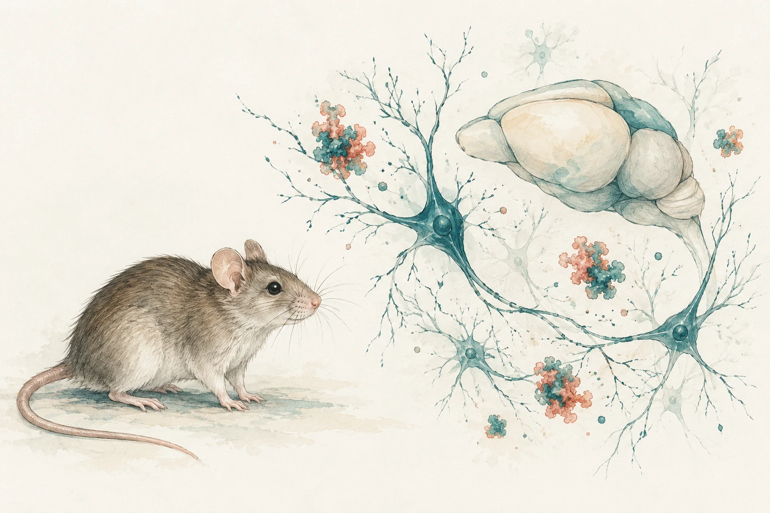
Ink and gouache illustration of a laboratory mouse beside a conceptual rodent brain, branching neurons and loose pigment clusters.
Mouse models · neurodegeneration

Model application

Neurodegeneration and proteinopathies

APP/PS1 amyloid models · transgenic or AAV-induced tauopathy

Region-specific assessment distinguishes amyloid or tau burden from the surrounding tissue response. Protein deposits, glial changes and neuronal integrity are related endpoints that need separate interpretation.

Endpoints, sampling and interpretation
Pathology endpoints
Regional plaque burden or phospho-tau labelling, with assessment of gliosis and neuronal integrity as required. The chosen stain, detection rules and tissue-area denominator are specified before measurement.
Sampling
Define brain regions, anatomical levels, model age and relevant controls. APP/PS1 amyloidosis and tauopathy remain distinct model examples. For AAV studies, consider transgene distribution and expression.
Interpretation
Deposit burden or marker intensity alone does not establish neuronal loss. Histology and independently defined neuronal endpoints provide the necessary tissue context.
Neuronal measurements and image analysis
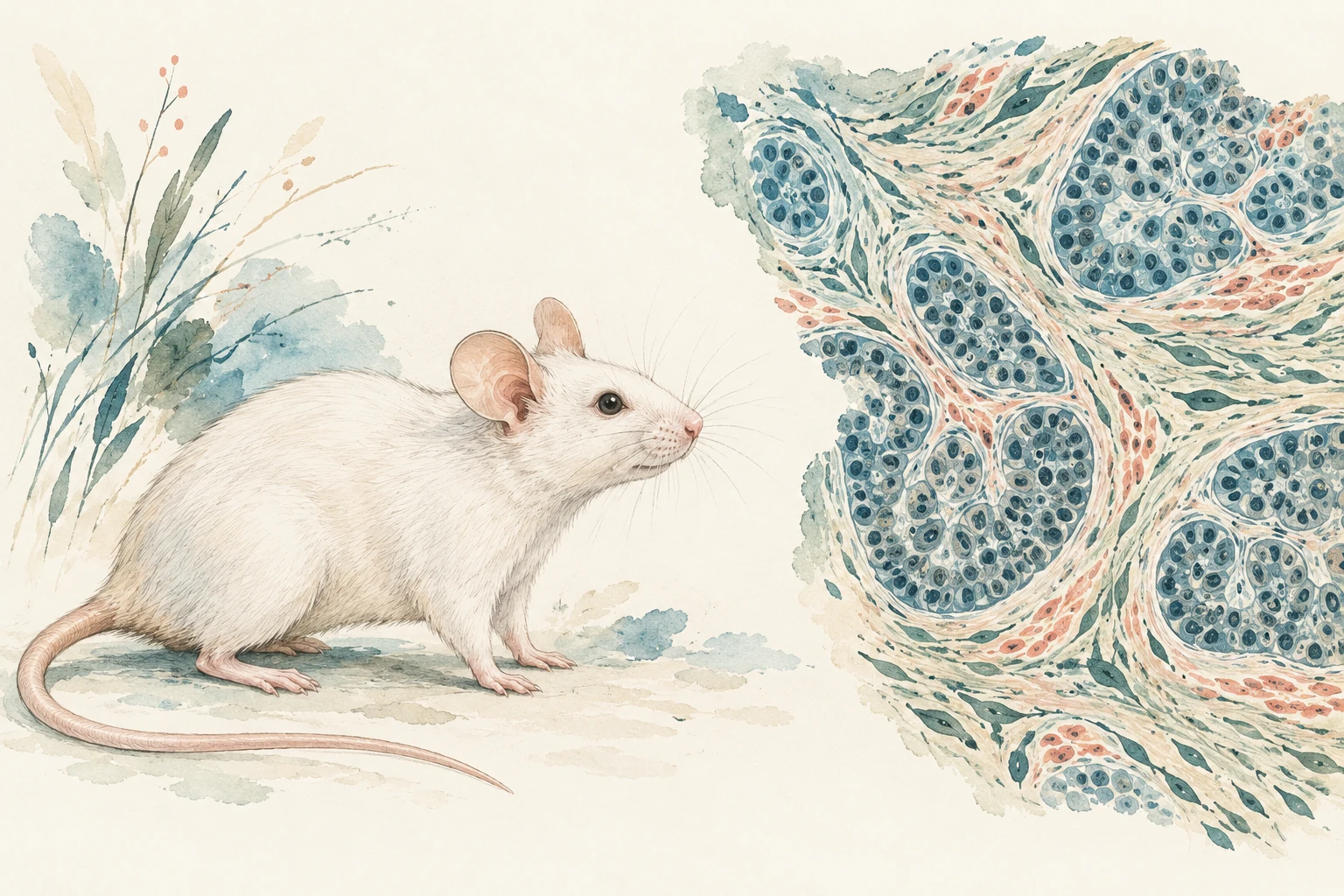
Editorial illustration of a laboratory mouse beside painted epithelial nests and surrounding stroma.

Model application

Cancer and immuno-oncology

Syngeneic tumours · xenografts and PDX

Histology adds tissue context to tumour-growth measurements. Viable tumour, necrosis, stroma and immune-cell distribution can be assessed in relation to the model, treatment mechanism and available biomarkers.

Endpoints, sampling and interpretation
Pathology endpoints
Viable and necrotic tumour area, stromal features, proliferation or other tissue biomarkers, and the location and density of selected immune-cell populations. Define regions and denominators before image analysis.
Sampling
Consider tumour heterogeneity, collection time and marker specificity. Syngeneic tumours in immunocompetent hosts and conventional human xenografts or PDX in immunodeficient hosts provide different immune contexts.
Interpretation
The model and host determine which immune findings can be interpreted. Tissue endpoints complement tumour-volume and other study data.
Oncology services and experience
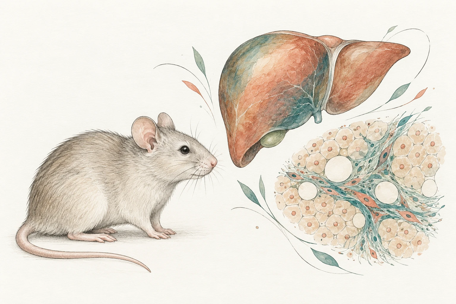
Editorial illustration of a laboratory mouse beside a separate liver silhouette and conceptual hepatic cells, lipid spaces and fibrous strands.
Mouse model · MASH and hepatic fibrosis

Model application

MASH and hepatic fibrosis

Diet-driven steatohepatitis · accelerated fibrosis models

Steatosis, inflammatory activity, hepatocellular injury and fibrosis describe different parts of the liver phenotype. Scoring and collagen measurements help characterise the response within the model’s metabolic and experimental context.

Endpoints, sampling and interpretation
Pathology endpoints
Steatosis, lobular inflammation, hepatocellular injury or ballooning where present, and the stage and distribution of fibrosis. Collagen-positive area can complement morphological assessment; it does not replace fibrosis stage.
Sampling
Use consistent liver-lobe and section sampling, with defined inclusion rules. Diet-driven and chemically accelerated models differ in their metabolic and injury context.
Interpretation
Less liver fat does not necessarily mean less injury or fibrosis. The components are reported separately and interpreted alongside the other study findings.
Histomorphometry within image analysis
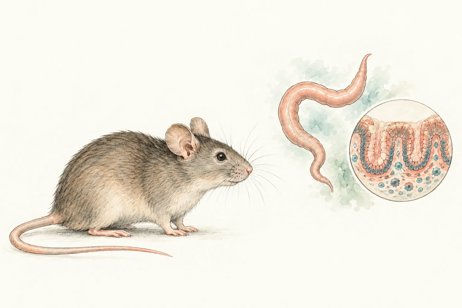
Editorial illustration of a laboratory mouse beside a separate colon and epithelial crypt vignette.

Model application

Inflammatory bowel disease

DSS-induced colitis in mice

Review of inflammation, epithelial injury and crypt architecture across the sampled colon. Defined scoring and measurements of injured or ulcerated mucosa describe both the severity and extent of the tissue changes.

Endpoints, sampling and interpretation
Pathology endpoints
Inflammatory infiltrates, erosion or ulceration, goblet-cell loss, crypt injury and regenerative changes. Injured or ulcerated mucosal length may be expressed relative to evaluable colon length.
Sampling
Check Swiss-roll completeness and orientation, and define the regions and time points to compare. Injury and repair can be unevenly distributed. Where the animal is the experimental unit, section-level outputs are summarised accordingly.
Interpretation
DSS is one model of intestinal injury and inflammation. The timing and model context matter when interpreting a change in score or ulcerated area.
My DSS-colitis experience

Before the first slide

Plan the tissue assessment.

We agree the biological question, tissue coverage, sampling, blinding, scoring and measurements. Image-analysis methods sit within that plan, with clear denominators and an experimental unit defined before summarising the data.

Scope an assessment

Related experience

My oligonucleotide safety work covers central and peripheral nervous-system assessment. Project examples include transgenic-rat β-amyloid assessment, intracerebral AAV studies in cynomolgus macaques, mdx muscular dystrophy and ex vivo human skin.

Contact

Discuss a study.

Email me the question, the material available and your key dates.