Services

Image analysis and computational pathology

I define tissue measurements, review image-analysis outputs and interpret them alongside the histology.

Applications across research and safety studies

Investigative studies

Fibrosis, inflammation, oncology and neural tissue assessment, alongside metabolic disease and organ-focused studies.

Toxicology

Lesion extent, cellular responses and recovery, with review of image quality and analysis outputs.

Application range and endpoints

Investigative studies

Applications include fibrosis and extracellular matrix, inflammation, oncology and the tumour microenvironment, tissue biomarkers, steatosis, neuroinflammation, demyelination and neurodegeneration, colitis, kidney, lung, cardiac and vascular disease, bone and cartilage, skin and wound healing, retinal disease, tissue localisation, organoids and other new approach methods, among others.

Toxicology

Lesion extent, hypertrophy, hyperplasia and atrophy, proliferation, apoptosis and regeneration, immune and haematopoietic changes, deposits, pigments and vacuolation, recovery assessments, lesion-detection assistance, annotation and evaluation, and image or specimen quality review.

Measurements may include area fractions, cell counts and densities, thickness, morphology, compartment-specific IHC/ISH signals and spatial relationships.

Explore the applications below, or see examples from my completed work.

Selected applications

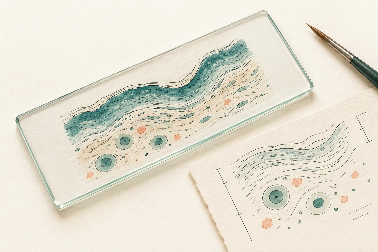
Ink and gouache illustration of flowing tissue-like ribbons and simple cell forms beside a glass slide.
Tissue structure and extracellular matrix

Image-analysis application

Fibrosis and tissue compartments

Collagen-positive area, tissue-compartment size and lesion distribution can be measured alongside the microscopic assessment of fibrosis. The region and denominator are chosen for the study question.

Measurement, review and interpretation
Measurement
Collagen-positive area as a fraction of the agreed evaluable tissue or compartment. Different regions can be reported separately where they represent different biological questions.
Sampling and review
Review section selection, staining, segmentation, normal structures and artefacts. Apply consistent inclusion rules and keep anatomical sampling comparable between animals.
Interpretation
Collagen area and fibrosis stage describe different features. Quantitative changes are interpreted with the architecture and distribution of the lesion.
Liver-fibrosis model applications
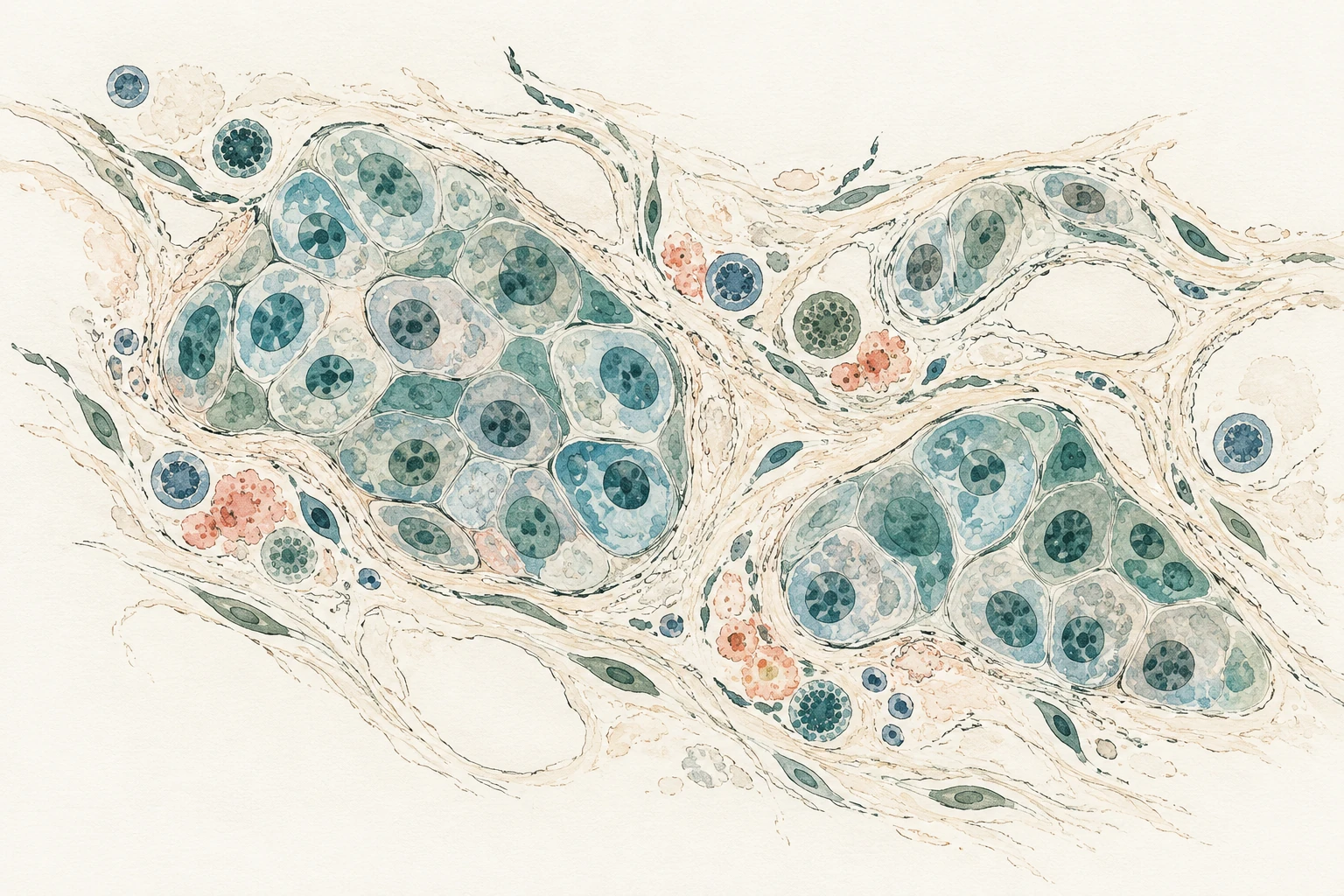
Ink and gouache illustration of drawn cell clusters, scattered cell-like forms and fine tissue-compartment boundaries.
Cells, tissue compartments and biomarkers

Image-analysis application

Oncology and tissue biomarkers

Positive-cell fractions, cell density and compartment-specific signals bring tissue context to biomarker measurements. Tumour/stroma boundaries and the location of immune-cell populations are part of the review.

My experience includes IHC/ISH, multiplex and fluorescence preparations, and collaboration with genomics scientists on spatial methods.

Measurement, review and interpretation
Measurement
Define the relevant cell population, staining compartment, positivity criteria and tissue region. Depending on the material, outputs may include positive-cell fractions, density, signal measurements or spatial relationships.
Sampling and review
Check cell detections, compartment boundaries, staining controls and thresholds. For multiplex material, consider channel quality and signal separation; for spatial comparisons, agree which cells and regions are being related.
Interpretation
A tissue-biomarker measurement needs its assay and study context. Signal intensity or spatial proximity alone does not establish biological function or predictive utility.
Preclinical oncology support
Editorial illustration of a laboratory rat beside a separate conceptual brain and cerebellar folia.
Rodent neuropathology

Image-analysis application

Neural tissue quantification

Regional cell counts, cell density and marker-positive area can complement assessment of neuronal injury, gliosis, myelin changes and protein deposits. Anatomical context remains central to the measurement.

Measurement, review and interpretation
Measurement
Define the cell population, anatomical region and denominator. Examples include cell counts per tissue area or layer length, and marker-positive area within a defined region.
Sampling and review
Agree section levels and review staining, tissue quality, layer boundaries and cell detections. Keep marker measurements distinct from independent assessment of neuronal integrity.
Interpretation
Reduced calbindin or other marker labelling does not by itself establish neuronal loss. Morphology and the relevant neuronal endpoints are interpreted together.
My collaborative Purkinje-cell work
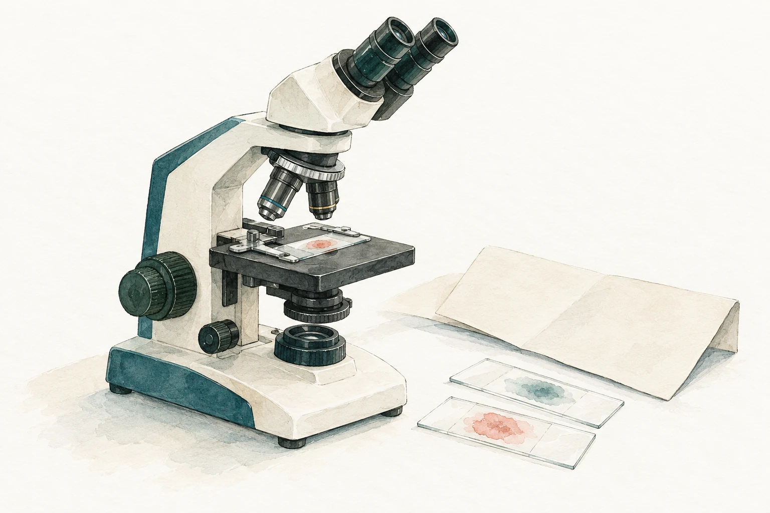
Ink and gouache illustration of a microscope, glass slides and paper in lagoon, forest and coral colours.
Pathology practice

Image-analysis application

Quantitative endpoints in toxicology

Lesion-area measurements, cellular changes and tissue-compartment measurements can support the interpretation of safety findings and recovery. Detection tools are reviewed against the underlying histology.

Measurement, review and interpretation
Measurement
Possible endpoints include lesion-area fractions, cell size or density, proliferative responses and selected tissue changes at treatment-end or recovery time points. Each lesion and evaluable tissue denominator are defined before analysis.
Sampling and review
Review overlapping findings, artefacts and unexpected detections. Assess method performance for the intended tissue, preparation and use. A slide-level classification or attention heatmap does not automatically provide a lesion-area measurement.
Interpretation
Quantitative outputs support the pathology interpretation. The GLP or non-GLP status, validation responsibility and quality arrangements are agreed for the image-analysis component.
Study-specific GLP and non-GLP arrangements
Editorial illustration of a glass slide and hand-drawn tissue boundaries with abstract measurement marks.

Image-analysis application

Annotation and algorithm review

I work with computational teams to define annotation criteria and review detections, segmentations and model outputs. The aim is to make the measurements and their limitations understandable in tissue terms.

Measurement, review and interpretation
Measurement
Reference annotations may identify tissue compartments, lesions or cell populations. The task, class definitions, ambiguous cases and expected output are agreed before annotation or review.
Sampling and review
Review representative material and difficult cases, with held-out test images where an algorithm is being evaluated. Examine errors by class and image context, including staining and scanner variation.
Interpretation
Performance is assessed within the intended scope. Annotation, output review, method validation and bespoke algorithm development are specified separately in the assignment.
Scope an image-analysis assignment

Image-analysis methods

Histomorphometry

Measuring tissue thickness, area, size and density. These examples combine pathologist-directed measurements or collaborative computational workflows with review of the histology.

Retinal thickness

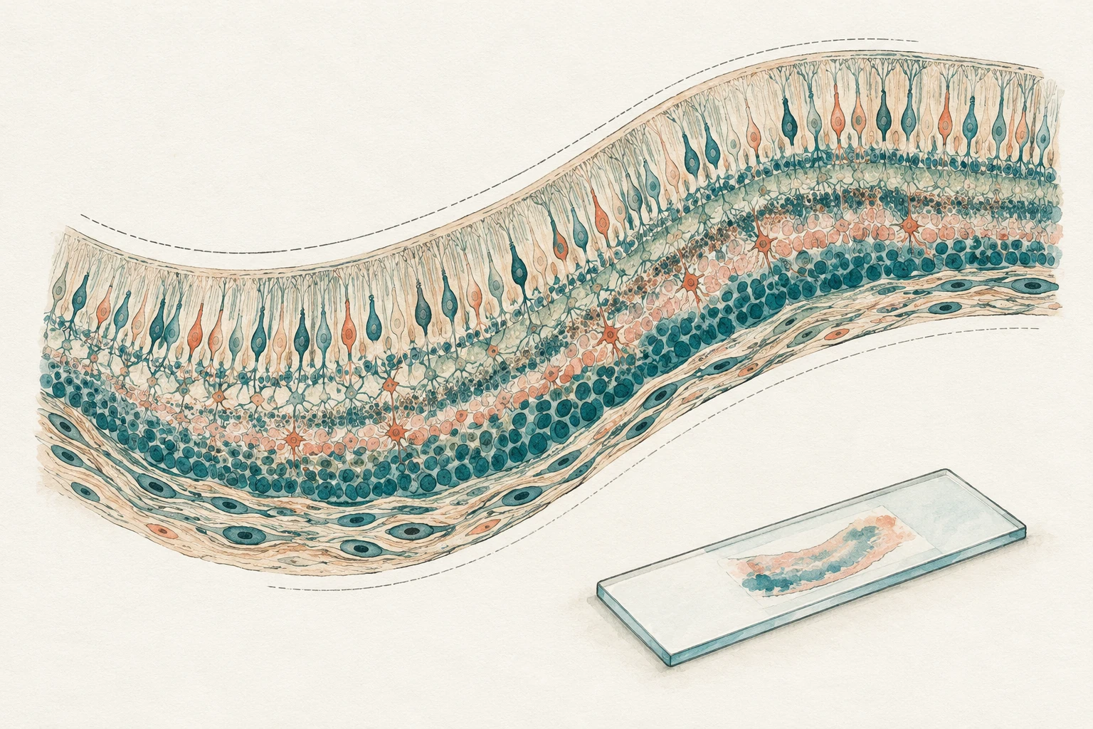
Hand-drawn layered retinal tissue concept with boundary traces and a glass microscope slide.

Thickness at defined anatomical locations on whole-slide images.

Sampling and review

Representative-section selection, consistent sampling, orientation and fixation or sectioning artefacts.

Skeletal-muscle morphometry

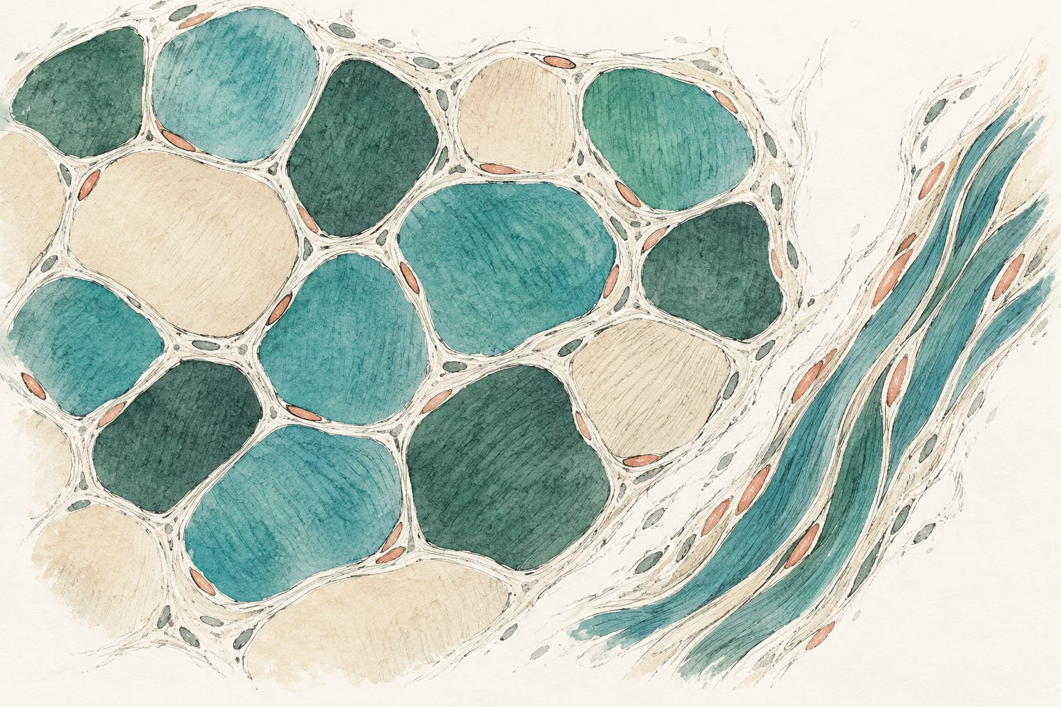
Ink and gouache illustration of rounded polygonal muscle-fibre-like profiles beside a flowing fibre bundle.

Myofiber-size measurements alongside assessment of necrosis, regeneration and inflammation, as in mdx muscular dystrophy work.

Sampling and review

The orientation of the section, the regions sampled and the pathological changes surrounding the measured fibers.

Adipose-tissue morphometry

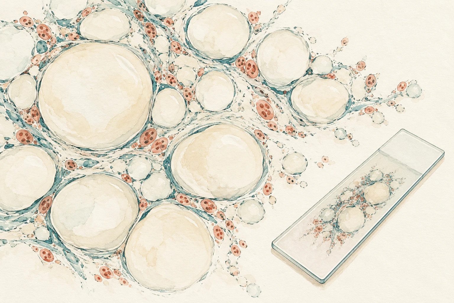
Ink and gouache illustration of rounded lipid-space-like profiles beside a microscope slide.

Image-derived lipid-space area and diameter in defined regions, with summaries at animal level where appropriate.

Sampling and review

Segmentation and sampling. The measurements describe two-dimensional profiles in sections, rather than three-dimensional cell volume.

Purkinje-cell density

Ink and gouache illustration of a single fan-like branching Purkinje neuron on ivory paper.

Purkinje-cell counts expressed per unit length of the Purkinje-cell layer. Collaborative work combined HALO AI segmentation and detection with QuPath processing.

Sampling and review

Tissue-layer boundaries, cell detections and the layer-length denominator, with pathologist review and correction of analysis outputs.

Define the measurement first.

We agree the sampled regions, denominator and experimental unit before analysis. Where the animal is the experimental unit, regional measurements can be summarised per animal.

Measurement choices and interpretation

The denominator may be tissue area or layer length. Section orientation, tissue quality and artefacts remain part of the review. Morphometry describes a measurement, such as retinal thickness; semiquantitative scoring describes a histological pattern, such as fibrosis severity.

Pathology and computation

Methods and working arrangements

I use QuPath for pathologist-directed measurements and have worked in collaborative HALO AI workflows. I also work with computational teams on reference annotations, output review and interpretation.

We agree the intended use, source images, sampling, analysis version and review criteria before work begins. The experimental unit and the way regional measurements are summarised are part of that plan.

Software, validation and quality arrangements

The software or code version, responsibility for method validation, data transfer and review records are specified for the assignment. The GLP or non-GLP status of an ancillary image-analysis component is agreed with the study team. Bespoke algorithm or software development requires a separate scope.

Inputs, scope and deliverables

What you receive

Measurements and reviewed outputs, interpreted in their tissue and study context.

  • An agreed annotation or measurement framework, with source images, analysis versions and documented outputs identified.
  • Review of representative detections, segmentations or measurements, including limitations.
  • A scientific interpretation connecting quantitative findings with the tissue and study context.
Planning the assignment
  • What is the intended use, biological question and tissue endpoint?
  • What images, sampling records, annotations and analysis outputs are available, and which software or code versions were used?
  • How are the experimental unit, aggregation, review criteria and responsibility for method validation defined?

Related services

Contact

Discuss a study.

Email me the question, the material available and your key dates.