
Methods guide · Neuropathology
Planning a neuropathology review for an oligonucleotide safety study
A useful neuropathology review starts with an agreed question and an identifiable set of tissues and records. This planning guide describes how I would scope a primary evaluation, independent peer review or focused consultation for an ASO or siRNA safety programme.
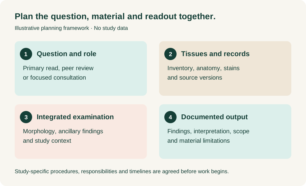
Define the role and question
Specify whether the assignment is a primary read, independent peer review or additional assessment of a particular finding. Define the question: for example, the distribution of a lesion, agreement on terminology and grading, or integration of routine histology with ancillary observations.
For a peer review, identify the study pathologist, the report and data versions, the groups and tissues to examine, and how discussions and any remaining differences will be documented. OECD and FDA guidance address the planning, management and documentation of GLP histopathology peer review; the study-specific arrangements remain essential.
Build the tissue and record inventory
List the species, treatment groups, dose and exposure information, administration route, treatment-end and recovery time points, and available clinical observations. Link each slide or whole-slide image to the animal, tissue, side, anatomical level and stain where recorded.
For a nervous-system assessment, agree the intended coverage of brain, spinal cord, dorsal root ganglia and peripheral nerves. Coverage should follow the study design and question; an available section does not establish that every relevant region was sampled. Record missing, damaged or unevaluable material and its effect on the conclusions.
Interpret morphology and ancillary findings together
Review routine histology for the nature, location, distribution and severity of changes. Consider image quality and section orientation before interpreting an apparent regional difference.
Additional stains can help address a defined question. Depending on the material and purpose, these may include markers such as IBA-1, GFAP or calbindin, or a Fluoro-Jade method. Their availability, performance and relationship to the examined H&E sections must be documented. A change in marker signal or an automated cell count does not, by itself, establish the underlying lesion, treatment relationship or adversity.
Agree the output and quality arrangements
The deliverable may be a pathology report, signed peer-review statement or focused scientific interpretation. It should identify what was examined, the methods and source versions, the findings and interpretation, and material limitations. Quantitative measurements require a defined region, denominator, sampling approach and experimental unit.
For GLP work, define applicable procedures, documentation, quality-assurance and record-retention arrangements with the responsible test facility and Study Director. Specify the status of each ancillary component. Digital examination also requires agreed image suitability, access and retention arrangements; FDA whole-slide imaging guidance is relevant where applicable.
What to send for an initial discussion
A study outline, proposed role, tissue and slide inventory, available stains, reporting requirements and target date are enough to begin. I offer flexible support for full evaluations and focused reviews; the scope and delivery schedule are agreed around the material and availability.
References and further reading
Discuss your study
Send the scientific question, available material and target dates. We can agree a focused review or a broader assignment around your study.